Types of Echo-cardiogram And What They Show
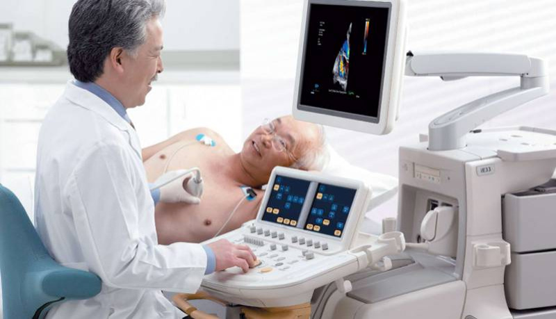
A Port Saint Lucie echocardiogram (echo) visually represents the beating heart. During an echocardiogram, your doctor will use ultrasonography to capture images of your heart’s valves and chambers by positioning a hand-held wand on your chest. The doctor can use the results to determine how well your heart pumps.
When assessing blood flow across the heart’s valves, doctors frequently use a combination of echo, Doppler ultrasound, and color Doppler. For an echocardiogram, no radiation is required. This distinguishes an echo from low-radiation diagnostic procedures like X-rays and CT scans.
How many distinct varieties of echocardiograms are there?
The echocardiogram might be of a few different varieties. When it comes to identifying and treating cardiac disease, each has its own set of advantages. Those things are:
- Exercise stress echocardiogram.
- Transthoracic echocardiogram.
- Transesophageal echocardiogram.
To what extent would I require an echocardiogram?
A physician may recommend an echo for a variety of reasons. Echocardiography may be necessary if:
- Your doctor is looking into your symptoms (either to diagnose a condition or to rule out plausible explanations).
- Your doctor has diagnosed you with cardiac illness. The echo is utilized for diagnosis and further investigation into the issue at hand.
- Your doctor wants to follow up on an existing diagnosis. People with valve problems, for instance, may benefit from routine echocardiograms.
- You are getting ready for an upcoming medical operation.
- Your doctor needs to follow up on a procedure or operation to see how it went.
Why is an echocardiography performed?
Many forms of heart illness are detectable by echocardiography. For example:
- Heart defects present at birth; congenital conditions.
- A disease of the heart muscle called cardiomyopathy.
- An infection of the heart’s lining or valves is called infectious endocarditis.
- Disease of the pericardial sac, the double-walled covering of the heart.
- Disease of the heart’s valves, the “doors” between the ventricles.
Alterations in your heart’s structure and function that an echo can detect may imply.
- Aneurysm of the aorta.
- Clotting occurs in the blood.
- A tumor in the heart.
Get echocardiography done today!
The structure and function of your heart can be assessed with great precision using echocardiography. If your provider suggests an echo, be sure you know what kind you’ll be getting and what to expect from it. For your doctor to receive a complete picture of your heart health, you may require more than one echo or a battery of tests using various methods.
In order to make sense of the images, you should probably ask your provider to break them down for you. You can gain confidence in your diagnosis and treatment if you actively participate in both.


